Published with this product (2)
2025Journal of Neuroimaging, 35(2), e70040
Phosphotungstic Acid Staining to Visualize the Vagus Nerve Perineurium Using Micro‐CT
Aniruddha R Upadhye, Eleana Cintron, Jichu Zhang, Jennifer Coleman, Chaitanya Kolluru, Michael W Jenkins, David Wilson, Nicole A Pelot, and Andrew J Shoffstall
doi:10.1111/jon.700402014The Journal of Neuroscience, 34(39): 13222-13233
Serena Quarta, Bastian E. Baeumer, Nadja Scherbakov, Manfred Andratsch, Stefan Rose-John, Georg Dechant, Christine E. Bandtlow, and Michaela Kress
doi:10.1523/JNEUROSCI.1209-13.2014
Description
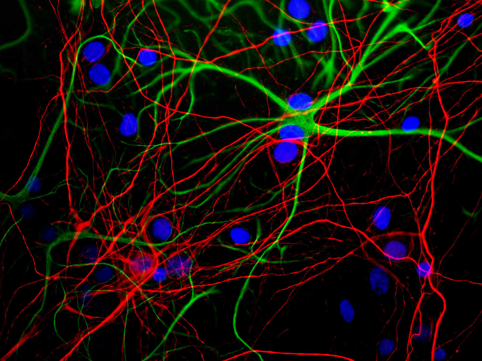
Neurofilaments can be defined as the intermediate or 10nm filaments found in specifically in neuronal cells. In the electron microscope, neurofilaments appear as 10nm diameter fibers of indeterminate length which generally have fine wispy protrusions from their sides. They are found particularly abundantly in axons of large projection neurons. Antibodies to the various neurofilament subunits are very useful cell type markers since the proteins are among the most abundant of the nervous system, are expressed only in neurons, and are biochemically very stable.
This antibody recognizes phosphorylated NF-H KSP (lysine-serine-proline) type sequences. In some species, there is some cross-reactivity with the related phosphorylated KSP sequences found in the related neurofilament subunit NF-M. The antibody recognizes NF-H strongly in all mammals tested to date and also in chicken. It recognizes neurofilaments in frozen sections in tissue culture and in formalin fixed sections
Related products
- α-Internexin/NF 66
- alpha Synuclein
- Artemin
- Bmi1
- CSF1 (Colony Stimulating Factor 1)
- FGF-2
- GAP43
- GAP43 (IgG)
- MAP2
- MAP2 (Microtubule assoc. protein 2)
- MAP2 (Microtubule Associated Protein 2)
- MARCKS (Myristoylated Alanine Rich C Kinase Substrate)
For research use only. Not for diagnostic or clinical use.