DEV — test website with test data. Orders here are not real and nothing is shipped or charged.

Tech support 866-350-1500

Pay by PO: enter it at checkout

Neuromics

Primary Antibody · Mouse IgG

Neurofilament NF-H

Species
Human, Mouse, Rat, Primate, Guinea Pig, Chicken, Rabbit, Bovine, Cat, Dog, Porcine, Equine
Applications
ICC, WB, IHC, IF

Description

Neurofilaments can be defined as the intermediate or 10nm filaments found in specifically in neuronal cells. In the electron microscope, neurofilaments appears as 10nm diameter fibres of indeterminate length which generally have fine wispy protrusions from their sides. They are found particularly abundantly in axons of large projection neurons. Antibodies to the various neurofilament subunits are very useful cell type markers since the proteins are among the most abundant of the nervous system, are expressed only in neurons, and are biochemically very stable.

Some studies have shown that levels of neurofilament heavy and neurofilament light are elevated in patients with Alzheimer's disease, frontotemporal lobe dementia, and vascular dementia. 
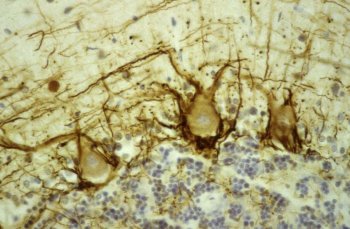
Human cerebellar cortex fixed in formalin, embedded in paraffin and stained with Mouse Monoclonal NF-H using the ABC (avidin biotin conjugate) method. The section was counterstained with heamatoxylin-eosin (blue). MO22120 stains prominent basket cell axons surrounding the large Purkinje neurons. Granule cell layer at bottom of image, molecular layer at top. Protocol on Data-sheet.
Rat spinal cord homogenate showing the major intermediate filament proteins of the nervous system (lane 1). The remaining lanes show blots of this material stainted with various antibodies including: Mouse Monoclonal NF-H (Lane 2), NF-M, NF-L, NF66 and GFAP. Protocol on Data-sheet.

Related products

For research use only. Not for diagnostic or clinical use.