Description
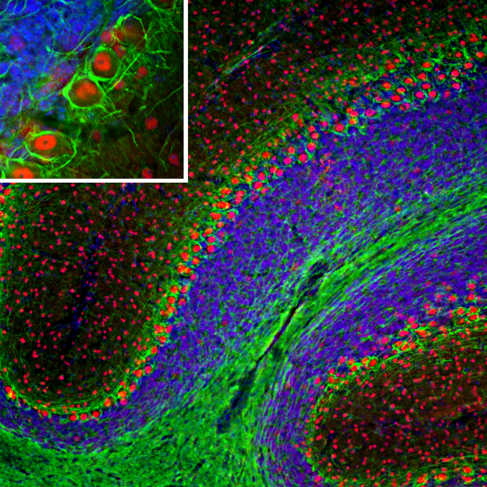
Neurofilaments are the 10nm or intermediate filament proteins found specifically in neurons, and are composed predominantly of three major proteins called NF-L, NF-M and NF-H, though other filament proteins may be included also. The major function of neurofilaments is likely to control the diameter of large axons. NF-L is the neurofilament light or low molecular weight polypeptide and runs on SDS-PAGE gels at 68-70kDa with some variability across species. Antibodies to NF-L like MCA-6H112 are useful for identifying neuronal cells and their processes in cell culture and sectioned material. NF-L antibody can also be useful for the visualization of neurofilament rich accumulations seen in many neurological diseases, such as Lou Gehrig’s disease (ALS), giant axon neuropathy, Charcot-Marie Tooth disease and others. Much interest has recently been focused on the detection of NF-L released from neurons into blood and CSF as a surrogate marker of primarily axonal loss in a variety of types of CNS injury and degeneration.
![Western blot analysis of different tissue lysates using mouse mAb to NF-L dilution 1:5,000 in green: [1] protein standard (red), [2] rat brain, [3] rat spinal cord, [4] mouse brain, and [5] mouse spinal cord. The strong band at 68kDa corresponds to the NF-L protein.](https://fxhiljpnygjqfppfgycj.supabase.co/storage/v1/object/public/catalog/products/MO22194/neuromics/images/02.jpg)
Related products
- α-Internexin/NF 66
- alpha Synuclein
- Artemin
- Bmi1
- CSF1 (Colony Stimulating Factor 1)
- FGF-2
- GAP43
- GAP43 (IgG)
- MAP2
- MAP2 (Microtubule assoc. protein 2)
- MAP2 (Microtubule Associated Protein 2)
- MARCKS (Myristoylated Alanine Rich C Kinase Substrate)
For research use only. Not for diagnostic or clinical use.