Description
LEF1 and TCF are members of the high mobility group (HMG) DNA binding protein family of transcription factors which consists of the following: Lymphoid enhancer factor 1 (LEF1), T Cell Factor 1 (TCF1), TCF3 and TCF4 (1). LEF1 and TCF1 were originally identified as important factors regulating early lymphoid development (2) and act downstream in Wnt signaling. LEF1/TCF bind to Wnt response elements to provide a docking site for β-catenin, which translocates to the nucleus to promote the transcription of target genes upon activation of Wnt signaling (3). LEF1/TCF proteins are dynamically expressed during development and aberrant activation of the Wnt signaling pathway is involved in many types of cancers including colon cancer (4,5).
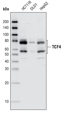
TCF4, also known as TCF7L2, is expressed widely during development. Gene targeting studies indicate that TCF4 is required to maintain the crypt stem cells of the small intestine (6,7). TCF4 has several splicing isoforms that are expressed differentially in tissues and during cancer progression (8,9). Studies also indicate that a variant of the TCF4 gene confers an increased risk of type 2 diabetes (10).
References: 1. Waterman, M.L. (2004) Cancer Metastasis Rev. 23, 41-52. 2. Schilham, M.W. and Clevers, H. (1998) Semin. Immunol. 10, 127-132. 3. Brantjes, H. et al. (2002) Biol. Chem. 383, 255-261. 4. Reya, T. and Clevers, H. (2005) Nature 434, 843-850. 5. Logan, C.Y. and Nusse, R. (2004) Annu. Rev. Cell Dev. Biol. 20, 781-810. 6. Cho, E.A. and Dressler, G.R. (1998) Mech. Dev. 77, 9-18. 7. Korinek, V. et al. (1998) Nat. Genet. 19, 379-383. 8. Howng, S.L. et al. (2004) Int. J. Oncol. 25, 1685-1692. 9. Shiina, H. et al. (2003) Clin. Cancer Res. 9, 2121-2132. 10. Grant, S.F. et al. (2006) Nat. Genet. 38, 320-323.
Related products
- ADAM15
- ALDH1L1
- Aldolase C
- Annexin A5
- Annexin A6
- AP-2 Alpha
- Aurora A Kinase
- Aurora A/B
- Aurora A/B Kinase
- Aurora B Kinase
- Bmi1
- BRAF35
For research use only. Not for diagnostic or clinical use.