Description
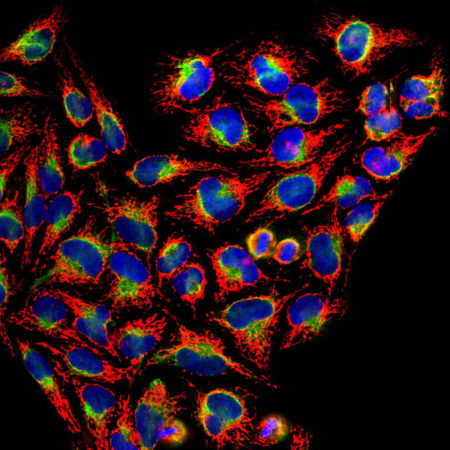
The heat shock proteins were discovered, as the name suggests, since they are heavily upregulated when cells are stressed by temperatures above the normal physiological range. They are expressed in unstressed cells also and have a normal function as chaperones, helping other proteins to fold correctly. The need for chaperones is much greater if a cell or tissue is stressed by heat, and so these proteins become heavily up regulated.. In addition to its role as a heat shock protein, HSP60 plays an important role in the transport and maintenance of mitochondrial proteins as well as the transmission and replication of mitochondrial DNA. HSP60 has also been implicated in the initiation and/or progression of some subtypes of cardiovascular disease (CVD).
Product Highlights:
![Western blot analysis of different tissue or cell lysates using rabbit pAb to HSP-60, RA22149, dilution 1:5,000 in red. [1] protein standard, [2] rat brain, [3] mouse brain, [4] NIH-3T3, [5] HEK293, [6] HeLa, [7] SH-SY5Y cells. The strong 60kDa band present in all preparations corresponds to HSP60 protein.](https://fxhiljpnygjqfppfgycj.supabase.co/storage/v1/object/public/catalog/products/RA22149/neuromics/images/02.jpg)
Related products
For research use only. Not for diagnostic or clinical use.