Description
mCherry is derived from proteins originally isolated from Cnidarians (jelly fish, sea anemones and corals), and is used as a fluorescent tracer in transfection and transgenic experiments. The prototype for these fluorescent proteins is Green Fluorescent Protein (GFP), which is a ~27kDa protein isolated originally from the jellyfish Aequoria victoria. The mCherry protein is derived from DsRed, a red fluorescent protein related to GFP isolated from so-called disc corals of the genus Discosoma. DsRed is similar in size and properties to GFP, but, obviously, produces a red rather than a green fluorochrome.
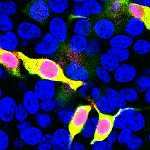
Several further cycles of mutation, directed modification and evolutionary selection produced mCherry, which has an excitation maximum at 587 nm and and emission maximum at 610 nm (4). We expressed the mCherry protein sequence shown in reference 4 in bacteria, purified out the mCherry and raised this rabbit polyclonal antibody. This was affinity purified and was found to stain a band of the expected size in HEK293 cells transfected with the pFin-EF1-mCherry vector designed to express mCherry. As shown below, the antibody does not stain any protein band in untransfected HEK293 cells.
Related products
- Fibrillarin
- Fibrillarin/Nop1p- Nucleolar Marker
- FluoGreen Tracer
- GFP
- Green Fluorescent Protein
- Green Fluorescent Protein (GFP)
- High-mobility group protein B1/HMGP1
- Lamin A/C
- mCherry
- mCherry - Mouse
- mCherry-Chicken
- Panspecific Nuclear Pore Complex Marker
For research use only. Not for diagnostic or clinical use.