Description
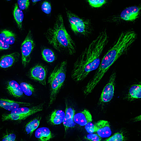
Fibrillarin is a highly conserved component of a nucleolar small ribonucleoprotein complex in mammals, involved in the processing of ribosomal RNA during ribosomal biogenesis. Fibrillarin was originally identified in humans since autoantibodies staining nucleoli were seen in some patients with the autoimmune disease scleroderma. Subsequently the protein fibrillarin was found to be the human homologue of Nop1p, a Saccharomyces cerevisiae nucleolar protein. Autoantibodies to fibrillarin are also seen in patients with the autoimmune disease systemic sclerosis.
RA22147 was made against recombinant human fibrillarin expressed in and purified from E. coli.
Product Highlights:
![Western blot analysis of cell lysates using rabbit pAb to fibrillarin, RA22147, dilution 1:5,000, in green: [1] protein standard (red), [2] NIH-3T3, [3] HEK293, [4] HeLa, [5] SH-SY5Y, [6] C6, and [7] COS1 cells. The strong band at ~35kDa corresponds to the fibrillarin protein.](https://fxhiljpnygjqfppfgycj.supabase.co/storage/v1/object/public/catalog/products/RA22147/neuromics/images/02.jpg)
Related products
- Fibrillarin
- Fibrillarin/Nop1p- Nucleolar Marker
- FluoGreen Tracer
- GFP
- Green Fluorescent Protein
- Green Fluorescent Protein (GFP)
- High-mobility group protein B1/HMGP1
- Lamin A/C
- mCherry
- mCherry - Mouse
- mCherry-Chicken
- mCherry-Rabbit
For research use only. Not for diagnostic or clinical use.